Understanding Hand Ultrasound: The Future of Diagnostic Imaging
Hand ultrasound is rapidly gaining recognition as a vital tool in the field of medical diagnostics. It provides a non-invasive, safe, and highly accurate imaging solution for various conditions affecting the hand and wrist. This article delves into the intricacies of hand ultrasound, discussing its technology, applications, benefits, and how it is revolutionizing patient care.
What is Hand Ultrasound?
Hand ultrasound refers to a specialized form of ultrasound imaging that focuses on the structures within the hand and wrist. Utilizing high-frequency sound waves, this technique captures real-time images of soft tissues, blood vessels, and even bones. The advent of portable ultrasound devices has made it increasingly accessible in various medical settings, allowing for immediate assessment and intervention.
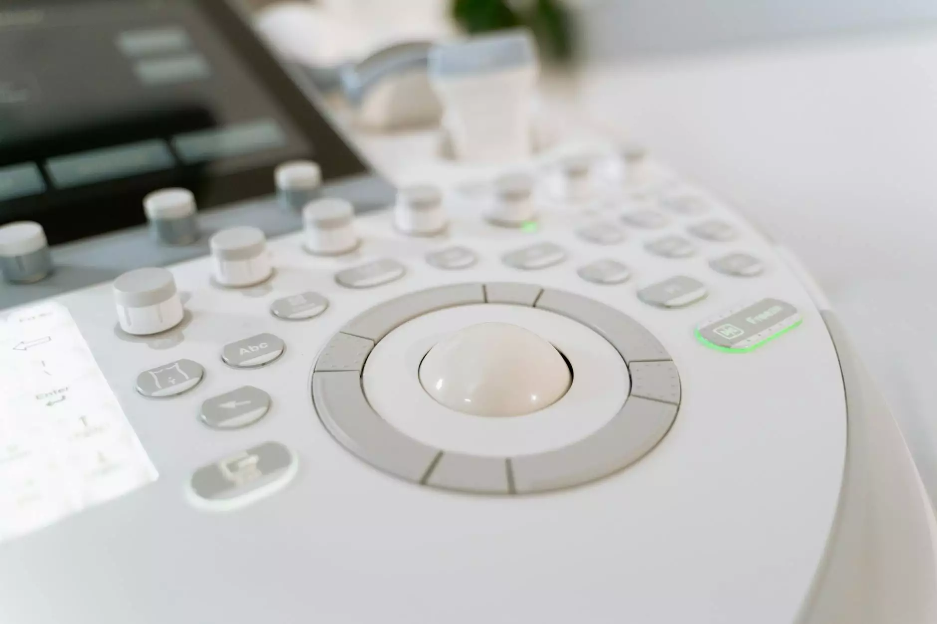
The Technology Behind Hand Ultrasound
The technology employs a transducer, which emits sound waves that bounce off tissues, creating images based on their density and composition. Here’s a brief overview of the hand ultrasound process:
- Preparation: The patient is often asked to position the hand in a specific manner, exposing the area of interest.
- Application of Gel: A water-based gel is applied to enhance sound wave transmission.
- Transducer Movement: The ultrasound technician moves the transducer across the skin, capturing real-time images.
- Image Interpretation: A radiologist or qualified healthcare provider analyzes the images for diagnostic purposes.
Key Benefits of Hand Ultrasound
The utilization of hand ultrasound offers numerous advantages that enhance diagnostic efficacy and patient experience. Here are some key benefits:
- Non-Invasive: Unlike other imaging techniques such as MRI or CT scans, hand ultrasound does not involve radiation or invasive procedures.
- Immediate Results: Ultrasound imaging can provide real-time feedback, allowing for timely clinical decisions.
- Cost-Effective: Hand ultrasound typically costs less than other imaging modalities, making it more accessible to patients.
- Portable Technology: The emergence of portable ultrasound units allows for examinations in various settings, from hospitals to clinics and even at home.
- High Sensitivity: Ultrasound is particularly effective at visualizing soft tissues, which is crucial in diagnosing conditions such as tendonitis or ligament tears.
Common Applications of Hand Ultrasound
Hand ultrasound is a versatile imaging technique used to diagnose a wide array of conditions. Some common applications include:
1. Assessment of Tendon Injuries
Tendons in the hand are susceptible to injuries, especially in athletes. Hand ultrasound can accurately visualize tendon tears, inflammation, and other pathological changes.
2. Evaluation of Ligament Damage
Ligament injuries, often resulting from trauma or repetitive strain, can be effectively assessed using this imaging technique.
3. Detection of Cysts or Tumors
Ultrasound can identify ganglion cysts or other masses within the hand that may require further investigation or intervention.
4. Blood Flow Assessment
Bearing importance in conditions like Raynaud's disease, hand ultrasound can evaluate blood flow and identify vascular abnormalities.
5. Guiding Injections
Ultrasound guidance can enhance the accuracy of injections in the hand, such as corticosteroids for inflammatory conditions.
Hand Ultrasound vs. Other Imaging Techniques
While hand ultrasound is exceptionally effective, it’s essential to understand how it compares to other imaging modalities:
1. Hand Ultrasound vs. MRI
While MRI provides detailed images of bones and soft tissues, it is more expensive, time-consuming, and requires specialized equipment. Hand ultrasound offers rapid results and is more comfortable for the patient.
2. Hand Ultrasound vs. X-rays
X-rays are excellent for visualizing bone fractures but fall short in assessing soft tissue injuries. Hand ultrasound excels in soft tissue imaging, making it invaluable for diagnosing tendon or ligament injuries.
Training and Proficiency in Hand Ultrasound
To maximize the benefits of hand ultrasound, it is crucial that the individuals performing these examinations possess the necessary training and competence. Here are some key points to consider:
- Specialized Training: Professionals should undergo specialized training to interpret ultrasound images accurately.
- Continued Education: As technology evolves, continuous learning is essential for staying updated with the latest advancements in ultrasound techniques.
- Multidisciplinary Approach: Collaboration among specialists, including rheumatologists, orthopedic surgeons, and dermatologists, can lead to comprehensive patient care.
Future Prospects of Hand Ultrasound
The future of hand ultrasound looks promising, with ongoing research and advancements in technology. Some potential developments include:
- AI Integration: The integration of artificial intelligence could enhance image analysis and diagnostic accuracy.
- Telemedicine Capabilities: Remote ultrasound imaging could facilitate consultations and diagnostics, particularly in underserved areas.
- Increased Portability: Innovations focusing on smaller, more user-friendly devices may make ultrasound accessible in even more settings.
Conclusion
In summary, hand ultrasound represents a significant advancement in diagnostic imaging, combining accessibility, safety, and efficacy. As technology continues to evolve and awareness grows, it is poised to become an indispensable tool in the assessment of hand and wrist conditions. Organizations like sonoscope.co.uk are at the forefront of this trend, providing essential services and education in the realm of ultrasound imaging.
As healthcare professionals and patients alike recognize the benefits of hand ultrasound, we can anticipate a future where this technology will play an even more pivotal role in enhancing healthcare outcomes.